Izzy Jayasinghe

image - SMS IJ Banner

Strong signals or crossed wires?

Electrical and chemical signals generated within cells, tissues and organ systems drive vital functions. Izzy Jayasinghe and her team investigate how these signals are relayed to trigger a heartbeat, and drive other bodily functions, by combing super-resolution microscopy tools they develop with existing technologies.

Most cells contain specialised hubs of signalling proteins and accessory molecules that propagate fast, large, and repeatable signals pivotal to healthy heart rhythm and other critical functions. Now that we know these hubs are wired differently in heart failure, chronic pain, and muscle weakness, Izzy’s team will build biochemical maps of these signals to unravel disease processes, and to design and refine novel precision therapeutics.

Learn more about the Signalling Nanodomains Lab

Cell signalling and disease

Rapid mobilization of ions or small molecules inside cells are amongst some of the fastest signalling mechanisms fundamental to life. They underpin physiologies such as the heartbeat, muscle contraction, neurotransmitter release, activation of gene transcription, and post-translational modifications. These mechanisms are also responsible for major human diseases and disabilities such as heart disease, cancer, paralysis, and chronic pain. Many such morbidities have no cure, whilst effectiveness of pharmacological treatments tends to be variable. At the core of some of the underpinning fast signals, are intracellular calcium signals (calcium sparks, puffs, waves, or transients) in excitable cells like muscle and neural tissues. They consist of specialised, nanoscale signalling domains that harbour ion channels and accessory molecules that coordinate to generate calcium sparks.

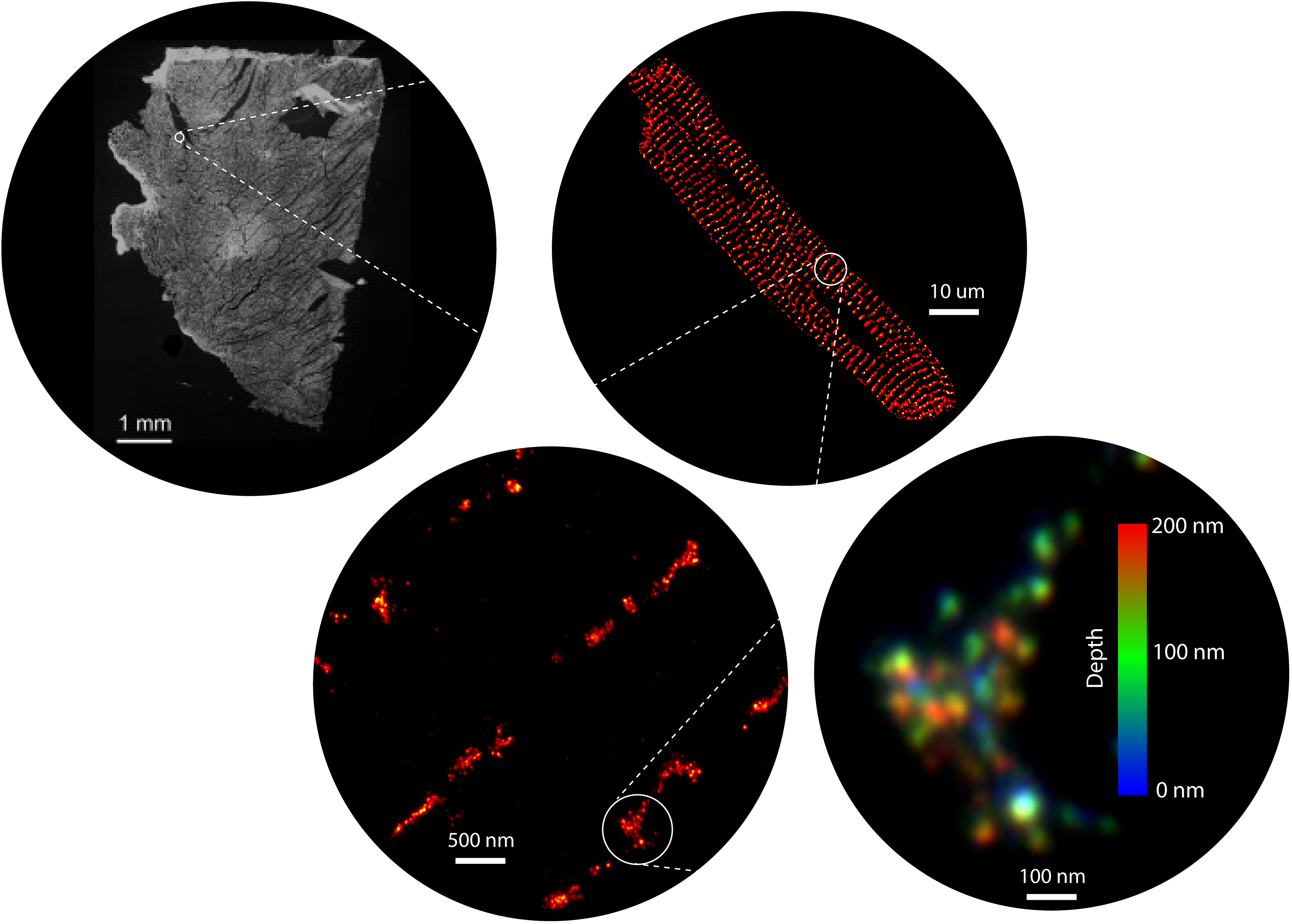
For over 15 years, our group have harnessed the power of a range of super-resolution microscopy technologies to resolve and visualise the shapes, locations, and molecular components intrinsic to these nanodomains (Jayasinghe et al, 2018). We now have the microscopy and analysis tools to not only map the position of each ion channel within these domains, but also to detect specific biochemical signatures on each channel, in situ (Sheard et al, 2019). For decades, the nanodomains and their signals have been imaged in isolation. However, one of our recent innovations, a correlative microscopy protocol (Hurley et al, 2021; Hurley et al, 2023), has allowed us to visualise the communications of channels such as ryanodine receptors can produce unexpected patterns of calcium signals. In particular, our discoveries have unearthed spatial heterogeneities in the organisation of calcium handling proteins of the myocardium. Our early observations suggest that these heterogeneities are not limited calcium handling proteins (such as ryanodine receptors (RyR2), L-type calcium channels, SERCA2A, and sodium calcium exchanger) but extend to regulatory molecules, second messengers, nucleic acids, and other structural proteins across the multitude of cells in the healthy heart. We also observe that these variabilities are accentuated in heart disease and may explain the limited effectiveness of the past and present generations of pharmacological therapies targeting the myocardial contractility.

Democratising super-resolution and high resolution optical imaging

Super-resolution microscopy, since its inception over two decades ago, has unlocked numerous secrets of the life processes. We have been one of the earliest adopters of this technology in its wide-ranging incarnations known commonly by numerous acronyms such as STORM, PALM, PAINT, STED and SIM. In spite of the nanometre-scale resolution that is on offer, access and usability of these methods remain modest due to the specialist nature of both the probes and the microscopy instrumentation. Over the past few years, our team have developed expertise in expansion microscopy (ExM) which parallels, and often exceeds, the resolution afforded through the traditional, localization- or optics-based super-resolution techniques. ExM uses molecular crosslinking and tissue clearing chemistries to obtain a three-dimensional imprint of cells, tissues, and/or whole organisms onto a polyacrylamide hydrogel (Sheard et al. 2019). These gels can then be osmotically swollen by a factor of >1000, effectively magnifying the intricate details of the sample that were previously too small to be resolved. ExM samples therefore allow us to visualise nanoscale features of cells and tissues conveniently with standard, and sometimes homebuilt, microscopes. Alongside of this methodology, we have been developing a series of tools that include high-throughput arrays, a new palette of fluorescent counterstains, distortion detection tools, gel automation robotics and 3D printable microscopy platforms to allow non-specialist microscopists to adopt super-resolution.

 

About Izzy Jayasinghe (she/her)

Associate Professor Izzy Jayasinghe was recruited to UNSW Sydney in 2023 as Head of the Department of Molecular Medicine in the School of Biomedical Sciences. Her research has and continues to be focus on developing new optical microscopy techniques for studying the organisation of the molecules of life, particularly proteins, within the heart. Izzy is a UKRI Future Leader Fellow at Sheffield University, where she is currently a Visiting Fellow and had served as Deputy Head of the Molecular and Cellular Biology Division since 2021. She built a track record in developing and applying new optical imaging methods during her postdoctoral fellowships in Queensland, and in Exeter (UK), aftre she completed a PhD in Auckland (New Zealand). In 2015, Izzy established her independent research program in the University of Leeds where she developed adaptations of optical imaging methods such as DNA-PAINT and Expansion Microscopy to study pathological nanoscale remodelling in the failing heart. Her current research focuses on developing more accessible, faster and higher resolution imaging methods for imaging optically-thick (and biologically more complex) samples. Izzy is a Fellow of the Royal Microscopical Society and advocates for Open Science and Equality and Inclusion in STEM.


Select Pubications

Seehra RS., Allouis, BHK, Sheard TMD, Spencer M, Shakespeare TB, Cadby A., Jayasinghe I. Geometry-preserving Expansion Microscopy microplates enable high fidelity nanoscale distortion mapping. Cell Reports Physical Science. 2023, DOI: 10.1016/j.xcrp.2023.101719

Hurley, M.E.H., White, E., Sheard, T.M.D., Kirton, H.M., Steele, D., Jayasinghe, I. Correlative nano-scale analysis of ryanodine receptor clusters at the origins of the elementary events of intracellular calcium signalling. Open Biology. 2023. 13; 230045.

Sheard, T.M.D., Hurley, M.E., White, E., Colyer, J., Norman, R., Pervolaraki, E., Narayanasamy, K., Hou, Y., Kirton, H., Yang, Z., Hunter, L., Shim, J., Clowsley, A.H., Smith, A.J., Baddeley, D., Soeller, C., Colman, M.A., Jayasinghe, I.; Three-dimensional and chemical mapping of intracellular signalling nanodomains in health and disease with enhanced expansion microscopy. ACS Nano. 2019. DOI: 10.1021/acsnano.8b08742 

Jayasinghe, I.*, Clowsley, A.H.*, Green, E., Harrison, C., Lutz, T.,Baddeley, D., di Michele, L., Soeller, C. True molecular scale visualisation of variable clustering properties of ryanodine receptors. Cell Reports. 2018; 22(2) 557-567. * Equal contributors 

Cully, T.R., Murphy, R.M., Roberts, L., Raastad, T., Fassett, R., Coombes, J.S., Jayasinghe, I.D., Launikonis, B.S. The plasmalemma of human skeletal muscle alters its structure to change its Ca2+ handling in response to heavy-load resistance exercise. Nature Communications. 2017; 8: 14266 

Full publication list


More Information

In the News


Career Highlights (select)

  • 2023 40 Under 40 Award, University of Auckland, New Zealand
  • 2022 Fellow of the Royal Microscopical Society, UK
  • 2021 Foundation Future Leader, Foundation of Science & Technology, London
  • 2020 Future Leader Fellow of UK Research & Innovation
  • 2018 Fellow of the Higher Education Academy, UK


Equity, Diversity & Inclusion Advocacy (select)

  • 2022 – Evidence presented to the Science & Technology Select Committee at the UK parliament about the lack of inclusion, and the lack of data measuring the diversity of women and LGBTQ+ people in STEM departments of UK universities. And Counter evidence presented to the Science & Technology Select Committee, against statements by government advisor, "There is a lot of hard maths in [physics] that I think that they would rather not do”. 
  • 2022 – The external advisor to working group conducting a review of Science Foundation Ireland's gender, equality, diversity and inclusion policies and practices.
  • 2019 – Co-author of open letter published in Sunday Times along with 1800 academic signatures affirming the work done by the Stonewall Diversity Champions program in UK universities. Full letter accessible here.


Projects

Our team consists of wide-ranging expertise ranging from biomedical/medical science, molecular biology, biophysics, bioengineering and chemistry. We are therefore open to Honours, PhD and postdoctoral researchers from a broad range of backgrounds. The following are a few examples of, but not limited to, some of the projects available with our group.

Uncovering the secret lives of regulatory molecules in the heart

The human myocardium is a highly heterogeneous tissue, comprising of a multitude of cell types such as fibroblasts, endothelial cells, and immune cells, in addition to the cardiomyocytes. Whilst the fundamental signalling mechanisms (e.g. excitation contraction coupling) are driven at the nanometre to micron scale, we now know that the signalling machinery is heterogeneously organised (Holmes et al. 2022). The expression, organisation, and modulation of the protein components of this machinery are all under intense regulation by a host of other regulatory molecules and nucleic acids (e.g. mRNAs and miRNAs) that are likely to be shared between the different cell types in the working myocardium. We hypothesise that the heterogeneities observed in the calcium handling proteins are a product of the heterogeneous expression and distribution of these molecular regulators. Owing to the lack of sensitive, multiplexed imaging techniques, this relationship has never been studied in the heart before. In this project, we will harness some of the compact antibody technologies to carry out multiplexed spatial proteomics combined with state-of-the-art localisation microscopy to map the spatial relationships between these two groups. We will initially develop engineered tissue assays that can help image this multi-cellular relationship, and then seek to apply the imaging pipeline to both human cardiac tissue biopsies and other mammalian models of heart failure.

Optical 3D imaging of the molecular polymorphisms of the giant ion channels in health and disease

For the first time, we will use optical microscopy to visualise the molecular structure of the largest ion channel in the body – the ryanodine receptor (RyR). At 2 MDa in mass, the tetrameric ryanodine receptor is the primary effector of the intracellular calcium signalling mechanisms that drive the heartbeat, musculoskeletal function, and neuronal excitability. RyRs of the heart (RyR2) were initially thought to be organised in highly regular array, however recent super-resolution imaging experiments have shown a more variable organisation (Jayasinghe et al, 2018). In addition, we have observed channel-to-channel variability of the regulatory partner proteins and post-translational modifications (e.g. site-specific phosphorylations). Whilst cryo-electron microscopy and crystallography have solved the molecular structure of purified RyR, the impacts of the variable partner interactions and chemical modifications on the overall tetrameric structure and conformations remain to be understood. Variable partner interactions and molecular modifications are thought to cause ‘de-tuning’ of the RyR arrays and heterogeneity in the efficacy in RyR-targeting drugs. We will combine high density fluorescent labelling and localisation microscopy to visualise the shapes and conformation of individual RyR2 in the presence of different partner proteins or phosphorylation conditions that mimic both healthy and disease physiologies. We will leverage state-of-the-art localisation microscopy platforms, 3D image visualisation tools to generate molecular maps of RyR2 at a level of both functional and structural detail never seen before.

Developing super-fast localisation microscopy

In this project, we will seek to advance the broadly used STochastic Optical Reconstruction Microscopy (STORM). In its classic implementation, STORM leverages aromatic fluorophores that can be switched reversibly between the fluorescent states and the longer-lasting dark states using reducing agents and high-powered laser illumination. This method however leads to steady photobleaching of fluorophores and accumulated phototoxicity of the sample over time which limits the length of the imaging time window available to the user and limits the quantitative nature of the images. Fluorescent nanodiamonds as STORM probes has emerged recently as an indestructible class of STORM probe alternatives to aromatic dyes. The spontaneous photoswitching that we have previously observed in nanodiamonds (Narayanasamy et al. 2020) is at least 5-times faster than that of regular STORM probes, and sensitive to the diamond surface chemistry. In this project, we will examine different surface chemistries applied to nanoscale diamonds and adopt them for developing targeted labels through conjugation with the new generations of monoclonal antibodies to visualise and track intracellular compartments such as endosomes and phagosomes. We will use state-of-the art localisation microscopy platforms and enhanced image analysis and reconstruction methods that leverage machine learning and algorithmic noise reduction.


Collaborators

Doan Ngo (Newcastle, Australia)
Aaron Sverdlov (Newcastle, Australia)
Robert Parton (Queensland, Australia)
Barbara Ciani (Sheffield, UK)
Philip Woodman (Manchester, UK)
Nikita Gamper (Leeds, UK)
Michael Colman (Leeds, UK)
Bradley Launikonis (Queensland, Australia)
Tom Gillingwater & Ross Jones (Edinburgh, UK)
Marie-Louise Ward (Auckland, New Zealand)
David Crossman (Auckland, New Zealand)
Melissa Mather (Nottingham, UK)
Andrea Gaion & Davide Sartori (ISPRA, Italy) 
John Colyer (Leeds, UK)