Felicity Davis is interested in how cells receive and decode information from their environment and communicate with each other to fulfil their biological functions.
The mammary gland changes dramatically during development, pregnancy, and once again when milk production is no longer needed. So, processes including cell death, tissue remodelling and regeneration can all be studied in mammary tissue.
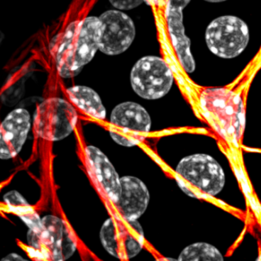
Felicity’s team uses live cell imaging to investigate calcium signals inside cells. Being able to image these signalling events across multiple scales lets them observe the behaviour of individual cells, and how this behaviour is coordinated with neighbouring cells to ultimately control the function of the organ.
How thousands to billions of cells are produced and arranged in space and time to execute a defined task at the cell level and cooperatively achieve a biological outcome at the organ level, remains an important question at the core of biomedical science. When an organ malfunctions, medical interventions typically focus on restoring function, where possible, or repairing or replacing damaged tissue. To optimally achieve these goals, however, we must first understand how the organ is formed, how it may regenerate and how it fundamentally functions. The lack of models and methods to visualise how cells in complex environments sense, decode and respond to developmental and physiological cues has limited progress in this area.
Using advanced microscopy—coupled with novel mouse models and computational platforms for image analysis—Felicity’s team is able to observe and quantify signal-response relationships in individual epithelial cells deep within living tissue. Her group is using this approach to provide new insights in to the molecular mechanisms that orchestrate the formation, function and failure of the mammary gland and other exocrine systems.
Learn more about the Calcium Signalling group.

